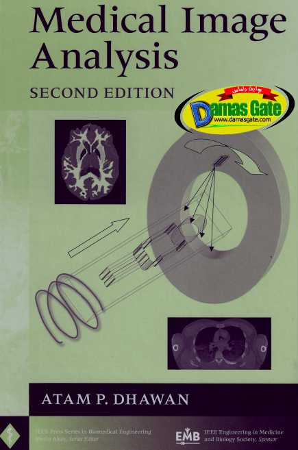
Medical Image Analysis - IEEE Biomedical Engineering Second Edition

Download
*

Description
-----------------------------------------------------------------------------------
Author(s): Atam P. Dhawan
Publisher: Wiley-IEEE Press
Date: 2011
Pages: 390
Format: PDF
Language: English
ISBN-10: 0470622059
ISBN-13: 978-0470622056
Size: 22.6 MB
-----------------------------------------------------------------------------------
Description:
-----------------------------------------------------------------------------------
Now updated - the most comprehensive reference of medical imaging modalities and image analysis techniques.
The last two decades have witnessed revolutionary advances in medical imaging and computerized medical image processing. With the advent and enhancement of numerous sophisticated medical imaging modalities, intelligent processing of multi-dimensional images has become critical in radiological and diagnostic applications.
This benchmark text takes a unique, all-inclusive approach to the topic - one that weaves together medical physics, medical imaging instrumentation, and advanced image analysis methods. This Second Edition is completely revised and expanded to provide a broader foundation, helping engineers, medical professionals, and students alike understand medical imaging principles, perform intelligent image interpretation, and navigate the intricacies of instrumentation, data collection, image reconstruction, and computerized image analysis for radiological computer-aided evaluation and diagnosis.
-----------------------------------------------------------------------------------
New chapters cover:
-----------------------------------------------------------------------------------
More in-depth description of recent developments in medical imaging instrumentation, including spiral CT, diagnostic ultrasound, functional MRI, and Diffusion Tension Imaging
Simultaneous multi-modality medical imaging, including CT-SPECT and CT-PET
Advanced medical image analysis and classification methods for computer-aided diagnosis, and therapeutic intervention
This updated edition presents individual chapters focused on x-ray, MRI, nuclear medicine, and ultrasound imaging modalities with additional details and recent advances. In addition, chapters on image reconstructions and visualizations have been significantly enhanced to include, respectively, 3-D statistical estimation-based reconstruction methods, feature classification and multi-modality image visualization. Examples with clinical images for medical image analysis and computer-aided diagnosis are provided throughout, as well as skill-building MATLAB exercises.
An ideal learning tool, this state-of-the-art resource can be used for one- or two-semester based senior undergraduate and/or graduate-level courses. Students in medical imaging and image processing, electrical and computer engineering, computer science, and biomedical engineering as well as physicians, medical physicists, and researchers will gain the knowledge to master the complexities of today's radiological and diagnostic applications.
-----------------------------------------------------------------------------------
Reviews:
-----------------------------------------------------------------------------------
This introductory text should be useful for senior undergraduate or graduate students in a medical image analysis course.
--Optical & Photonics News, April 2006
...an excellent introductory text.... I would strongly recommend the text... a welcomed addition to the personal libraries of students within these professional disciplines.
--Health Physics, Vol. 86, No. 4, March 2004
The book clearly reviews the various basic elements of medical imaging and of imaging reconstruction, and I would recommend it for the library of any Department of Radiology to be read by the junior residents and also consulted by the more senior members.
--Clinical Imaging, Vol. 28, No. 1 January/February 2004
-----------------------------------------------------------------------------------
About the Author:
-----------------------------------------------------------------------------------
Atam P. Dhawan, PhD, is Distinguished Professor in the Electrical and Computer Engineering Department at New Jersey Institute of Technology. He teaches courses in biomedical engineering and has supervised approximately fifty graduate students, including twenty-one PhD students.
Dr. Dhawan is a Fellow of the IEEE and the recipient of numerous national and international awards. He has published more than 200 research articles in refereed journals, conference proceedings, and edited books.
Dr. Dhawan has chaired numerous study sections and review panels for the National Institutes of Health in biomedical computing and medical imaging and health informatics. His current research interests are medical imaging, multi-modality medical image analysis, multi-grid image reconstruction, wavelets, genetic algorithms, neural networks, adaptive learning, and pattern recognition.
-----------------------------------------------------------------------------------
PREFACE TO THE SECOND EDITION
Radiological sciences in the last two decades have witnessed a revolutionary prog-
ress in medical imaging and computerized medical image processing. The develop-
ment and advances in multidimensional medical imaging modalities such as X-ray mammography, X-ray computed tomography (X-ray CT), single photon computed tomography (SPECT), positron emission tomography (PET), ultrasound, magnetic
resonance imaging (MRI), and functional magnetic resonance imaging (fMRI) have provided important radiological tools in disease diagnosis, treatment evaluation, and intervention for significant improvement in health care. The development of imaging instrumentation has inspired the evolution of new computerized methods of image reconstruction, processing, and analysis for better understanding and interpretation of medical images. hnage processing and analysis methods have been used to help physicians to make important medical decisions through physician-computer inter-
action. Recently, intelligent or model-based quantitative image analysis approaches have been explored for computer-aided diagnosis to improve the sensitivity and specificity of radiological tests involving medical images.
Medical imaging in diagnostic radiology has evolved as a result of the significant contributions of a number of disciplines from basic sciences, engineering, and medi-
cine. Computerized image reconstruction, processing, and analysis methods have een developed for medical imaging applications. The application-domain knowl-
dge has been used in developing models for accurate analysis and interpretation.
-----------------------------------------------------------------------------------
Author(s): Atam P. Dhawan
Publisher: Wiley-IEEE Press
Date: 2011
Pages: 390
Format: PDF
Language: English
ISBN-10: 0470622059
ISBN-13: 978-0470622056
Size: 22.6 MB
-----------------------------------------------------------------------------------
Description:
-----------------------------------------------------------------------------------
Now updated - the most comprehensive reference of medical imaging modalities and image analysis techniques.
The last two decades have witnessed revolutionary advances in medical imaging and computerized medical image processing. With the advent and enhancement of numerous sophisticated medical imaging modalities, intelligent processing of multi-dimensional images has become critical in radiological and diagnostic applications.
This benchmark text takes a unique, all-inclusive approach to the topic - one that weaves together medical physics, medical imaging instrumentation, and advanced image analysis methods. This Second Edition is completely revised and expanded to provide a broader foundation, helping engineers, medical professionals, and students alike understand medical imaging principles, perform intelligent image interpretation, and navigate the intricacies of instrumentation, data collection, image reconstruction, and computerized image analysis for radiological computer-aided evaluation and diagnosis.
-----------------------------------------------------------------------------------
New chapters cover:
-----------------------------------------------------------------------------------
More in-depth description of recent developments in medical imaging instrumentation, including spiral CT, diagnostic ultrasound, functional MRI, and Diffusion Tension Imaging
Simultaneous multi-modality medical imaging, including CT-SPECT and CT-PET
Advanced medical image analysis and classification methods for computer-aided diagnosis, and therapeutic intervention
This updated edition presents individual chapters focused on x-ray, MRI, nuclear medicine, and ultrasound imaging modalities with additional details and recent advances. In addition, chapters on image reconstructions and visualizations have been significantly enhanced to include, respectively, 3-D statistical estimation-based reconstruction methods, feature classification and multi-modality image visualization. Examples with clinical images for medical image analysis and computer-aided diagnosis are provided throughout, as well as skill-building MATLAB exercises.
An ideal learning tool, this state-of-the-art resource can be used for one- or two-semester based senior undergraduate and/or graduate-level courses. Students in medical imaging and image processing, electrical and computer engineering, computer science, and biomedical engineering as well as physicians, medical physicists, and researchers will gain the knowledge to master the complexities of today's radiological and diagnostic applications.
-----------------------------------------------------------------------------------
Reviews:
-----------------------------------------------------------------------------------
This introductory text should be useful for senior undergraduate or graduate students in a medical image analysis course.
--Optical & Photonics News, April 2006
...an excellent introductory text.... I would strongly recommend the text... a welcomed addition to the personal libraries of students within these professional disciplines.
--Health Physics, Vol. 86, No. 4, March 2004
The book clearly reviews the various basic elements of medical imaging and of imaging reconstruction, and I would recommend it for the library of any Department of Radiology to be read by the junior residents and also consulted by the more senior members.
--Clinical Imaging, Vol. 28, No. 1 January/February 2004
-----------------------------------------------------------------------------------
About the Author:
-----------------------------------------------------------------------------------
Atam P. Dhawan, PhD, is Distinguished Professor in the Electrical and Computer Engineering Department at New Jersey Institute of Technology. He teaches courses in biomedical engineering and has supervised approximately fifty graduate students, including twenty-one PhD students.
Dr. Dhawan is a Fellow of the IEEE and the recipient of numerous national and international awards. He has published more than 200 research articles in refereed journals, conference proceedings, and edited books.
Dr. Dhawan has chaired numerous study sections and review panels for the National Institutes of Health in biomedical computing and medical imaging and health informatics. His current research interests are medical imaging, multi-modality medical image analysis, multi-grid image reconstruction, wavelets, genetic algorithms, neural networks, adaptive learning, and pattern recognition.
-----------------------------------------------------------------------------------
PREFACE TO THE SECOND EDITION
Radiological sciences in the last two decades have witnessed a revolutionary prog-
ress in medical imaging and computerized medical image processing. The develop-
ment and advances in multidimensional medical imaging modalities such as X-ray mammography, X-ray computed tomography (X-ray CT), single photon computed tomography (SPECT), positron emission tomography (PET), ultrasound, magnetic
resonance imaging (MRI), and functional magnetic resonance imaging (fMRI) have provided important radiological tools in disease diagnosis, treatment evaluation, and intervention for significant improvement in health care. The development of imaging instrumentation has inspired the evolution of new computerized methods of image reconstruction, processing, and analysis for better understanding and interpretation of medical images. hnage processing and analysis methods have been used to help physicians to make important medical decisions through physician-computer inter-
action. Recently, intelligent or model-based quantitative image analysis approaches have been explored for computer-aided diagnosis to improve the sensitivity and specificity of radiological tests involving medical images.
Medical imaging in diagnostic radiology has evolved as a result of the significant contributions of a number of disciplines from basic sciences, engineering, and medi-
cine. Computerized image reconstruction, processing, and analysis methods have een developed for medical imaging applications. The application-domain knowl-
dge has been used in developing models for accurate analysis and interpretation.
Download
*