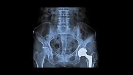
How To Read Hip And Pelvis X-Ray
Comprehensive Hip and Pelvis Radiology Course"
What you'll learn
*Interpret hip and pelvis X-rays* accurately and confidently.
Recognize normal anatomy* and variants.
Identify traumatic conditions*, fractures, and dislocations.
Diagnose degenerative and inflammatory conditions*, such as osteoarthritis and rheumatoid arthritis.
Understand hip arthroplasty* complications and imaging.
Develop skills* in X-ray interpretation, diagnosis, and patient management.
Apply knowledge* in clinical practice,

Requirements
Basic knowledge of human anatomy*: Familiarity with medical terminology and basic anatomy.
Medical background*: Radiologists, orthopedic surgeons, medical students, and healthcare professionals
Basic understanding of X-ray imaging principles.
Technical Requirements 1. Computer or laptop. 2. Internet connection. 3. Udemy account.
Description
Hip and Pelvis Radiology X-ray Enhance your diagnostic skills and improve patient outcomes with this comprehensive online course.Course OverviewThis extensive course covers the fundamentals and advanced concepts of hip and pelvis radiology, empowering healthcare professionals to accurately interpret X-rays, diagnose traumatic and degenerative conditions, and provide optimal patient care.
What You'll Learn
1. *Hip and pelvis anatomy*: Understand normal anatomy, variants, and developmental conditions.2. *Traumatic conditions*: Identify fractures, dislocations, and soft tissue injuries.3. *Degenerative diseases*: Diagnose osteoarthritis, rheumatoid arthritis, and other conditions.4. *Hip arthroplasty*: Understand complications, imaging techniques, and post-operative evaluation.5. *Imaging techniques*: Master AP, lateral, Judet, and oblique views.Key Features1. *67 in-depth lectures* with high-quality images and diagrams.2. *Interactive quizzes* to reinforce learning.3. *Downloadable resources* for future reference.4. *Certificate of completion* upon finishing the course.Target Audience1. Radiologists2. Orthopedic surgeons3. Medical students4. Healthcare professionals seeking continuing educationWhat You'll Achieve1. Enhanced diagnostic skills2. Improved patient outcomes3. Confidence in interpreting hip and pelvis X-rays4. Advanced knowledge of traumatic and degenerative conditionsRequirements1. Basic knowledge of human anatomy2. Medical background (recommended)3. English proficiencyStudents will learn:1. *Interpret hip and pelvis X-rays* accurately and confidently.2. *Recognize normal anatomy* and variants.3. *Identify traumatic conditions*, fractures, and dislocations.4. *Diagnose degenerative and inflammatory conditions*, such as osteoarthritis and rheumatoid arthritis.5. *Understand hip arthroplasty* complications and imaging.6. *Develop skills* in X-ray interpretation, diagnosis, and patient management.7. *Apply knowledge* in clinical practice, enhancing patient care
Overview
Section 1: Introduction
Lecture 1 Introduction
Section 2: Radiographic views
Lecture 2 AP view
Lecture 3 Lateral view
Lecture 4 Frog legs view
Lecture 5 Inlet and outlet AP view
Lecture 6 Judet view
Lecture 7 Obturator oblique
Lecture 8 Iliac oblique
Lecture 9 Femur view
Section 3: Pelvis &Hip anatomy
Lecture 10 Anatomy 1
Lecture 11 Anatomy 2
Lecture 12 Anatomy 3
Lecture 13 Anatomy 4
Lecture 14 Blood supply
Lecture 15 Anatomy 6
Lecture 16 Anatomy 7
Lecture 17 Anatomy 8
Lecture 18 Anatomy 9
Lecture 19 Proximal femur
Lecture 20 Developing skeleton
Lecture 21 Anatomy 12
Lecture 22 Anatomy 13
Section 4: Hip variants
Lecture 23 Ischiopubic synchondrosis
Lecture 24 Os acetabulum
Lecture 25 Synovial pit
Lecture 26 Pseudo lesion
Section 5: Trauma in hip and pelvis
Lecture 27 Traumatic conditions
Lecture 28 Normal apophysis
Lecture 29 Classification of pelvic fracture
Lecture 30 Main bone ring fracture
Lecture 31 Straddle fracture
Lecture 32 Bucket handle
Lecture 33 Malgagne fracture
Lecture 34 Open book #
Lecture 35 Acetabulum #
Lecture 36 Sacral#
Lecture 37 Pubic #
Lecture 38 Apophysis #
Lecture 39 Checklist
Lecture 40 Proximal femur #
Lecture 41 Intra capsular
Lecture 42 Garden types
Lecture 43 Extra capsular
Lecture 44 Hip dislocation
Lecture 45 THR dislocation
Lecture 46 Anterior hip dislocation
Lecture 47 Central hip dislocation
Lecture 48 Examples of dislocation
Section 6: Non traumatic pelvis and hip
Lecture 49 Coxa arthrosis
Lecture 50 Rheumatoid
Lecture 51 T. B
Lecture 52 AVN
Lecture 53 Looser zones
Lecture 54 Banana#
Lecture 55 Avulsion lesser trochanteric #
Lecture 56 Protrusion acetabulum
Lecture 57 Coxa magna
Lecture 58 Coxa plana
Lecture 59 Coxa Valga
Lecture 60 Coxa vara
Lecture 61 FAI
Lecture 62 Cam type
Lecture 63 Pincer type
Lecture 64 Hip arthroplasty
Lecture 65 Peri prothesis fracture
Lecture 66 Luxation in hip arthroplasty
Lecture 0 Quiez answers
This course is ideal for: Medical Professionals 1. Radiologists 2. Orthopedic surgeons 3. Medical students 4. Residents and fellows 5. Healthcare professionals Allied Health Professionals 1. Physician assistants 2. Nurse practitioners 3. Radiologic technologists 4. Orthopedic nurses Specialized Audiences 1. Musculoskeletal radiologists 2. Orthopedic specialists 3. Sports medicine professionals 4. Physical medicine and rehabilitation specialists Anyone seeking continuing education or professional development in hip and pelvis radiology.
Published 12/2024
MP4 | Video: h264, 1920x1080 | Audio: AAC, 44.1 KHz
Language: English | Size: 259 MB | Duration: 2h 9m
Download
*
Comprehensive Hip and Pelvis Radiology Course"
What you'll learn
*Interpret hip and pelvis X-rays* accurately and confidently.
Recognize normal anatomy* and variants.
Identify traumatic conditions*, fractures, and dislocations.
Diagnose degenerative and inflammatory conditions*, such as osteoarthritis and rheumatoid arthritis.
Understand hip arthroplasty* complications and imaging.
Develop skills* in X-ray interpretation, diagnosis, and patient management.
Apply knowledge* in clinical practice,

Requirements
Basic knowledge of human anatomy*: Familiarity with medical terminology and basic anatomy.
Medical background*: Radiologists, orthopedic surgeons, medical students, and healthcare professionals
Basic understanding of X-ray imaging principles.
Technical Requirements 1. Computer or laptop. 2. Internet connection. 3. Udemy account.
Description
Hip and Pelvis Radiology X-ray Enhance your diagnostic skills and improve patient outcomes with this comprehensive online course.Course OverviewThis extensive course covers the fundamentals and advanced concepts of hip and pelvis radiology, empowering healthcare professionals to accurately interpret X-rays, diagnose traumatic and degenerative conditions, and provide optimal patient care.
What You'll Learn
1. *Hip and pelvis anatomy*: Understand normal anatomy, variants, and developmental conditions.2. *Traumatic conditions*: Identify fractures, dislocations, and soft tissue injuries.3. *Degenerative diseases*: Diagnose osteoarthritis, rheumatoid arthritis, and other conditions.4. *Hip arthroplasty*: Understand complications, imaging techniques, and post-operative evaluation.5. *Imaging techniques*: Master AP, lateral, Judet, and oblique views.Key Features1. *67 in-depth lectures* with high-quality images and diagrams.2. *Interactive quizzes* to reinforce learning.3. *Downloadable resources* for future reference.4. *Certificate of completion* upon finishing the course.Target Audience1. Radiologists2. Orthopedic surgeons3. Medical students4. Healthcare professionals seeking continuing educationWhat You'll Achieve1. Enhanced diagnostic skills2. Improved patient outcomes3. Confidence in interpreting hip and pelvis X-rays4. Advanced knowledge of traumatic and degenerative conditionsRequirements1. Basic knowledge of human anatomy2. Medical background (recommended)3. English proficiencyStudents will learn:1. *Interpret hip and pelvis X-rays* accurately and confidently.2. *Recognize normal anatomy* and variants.3. *Identify traumatic conditions*, fractures, and dislocations.4. *Diagnose degenerative and inflammatory conditions*, such as osteoarthritis and rheumatoid arthritis.5. *Understand hip arthroplasty* complications and imaging.6. *Develop skills* in X-ray interpretation, diagnosis, and patient management.7. *Apply knowledge* in clinical practice, enhancing patient care
Overview
Section 1: Introduction
Lecture 1 Introduction
Section 2: Radiographic views
Lecture 2 AP view
Lecture 3 Lateral view
Lecture 4 Frog legs view
Lecture 5 Inlet and outlet AP view
Lecture 6 Judet view
Lecture 7 Obturator oblique
Lecture 8 Iliac oblique
Lecture 9 Femur view
Section 3: Pelvis &Hip anatomy
Lecture 10 Anatomy 1
Lecture 11 Anatomy 2
Lecture 12 Anatomy 3
Lecture 13 Anatomy 4
Lecture 14 Blood supply
Lecture 15 Anatomy 6
Lecture 16 Anatomy 7
Lecture 17 Anatomy 8
Lecture 18 Anatomy 9
Lecture 19 Proximal femur
Lecture 20 Developing skeleton
Lecture 21 Anatomy 12
Lecture 22 Anatomy 13
Section 4: Hip variants
Lecture 23 Ischiopubic synchondrosis
Lecture 24 Os acetabulum
Lecture 25 Synovial pit
Lecture 26 Pseudo lesion
Section 5: Trauma in hip and pelvis
Lecture 27 Traumatic conditions
Lecture 28 Normal apophysis
Lecture 29 Classification of pelvic fracture
Lecture 30 Main bone ring fracture
Lecture 31 Straddle fracture
Lecture 32 Bucket handle
Lecture 33 Malgagne fracture
Lecture 34 Open book #
Lecture 35 Acetabulum #
Lecture 36 Sacral#
Lecture 37 Pubic #
Lecture 38 Apophysis #
Lecture 39 Checklist
Lecture 40 Proximal femur #
Lecture 41 Intra capsular
Lecture 42 Garden types
Lecture 43 Extra capsular
Lecture 44 Hip dislocation
Lecture 45 THR dislocation
Lecture 46 Anterior hip dislocation
Lecture 47 Central hip dislocation
Lecture 48 Examples of dislocation
Section 6: Non traumatic pelvis and hip
Lecture 49 Coxa arthrosis
Lecture 50 Rheumatoid
Lecture 51 T. B
Lecture 52 AVN
Lecture 53 Looser zones
Lecture 54 Banana#
Lecture 55 Avulsion lesser trochanteric #
Lecture 56 Protrusion acetabulum
Lecture 57 Coxa magna
Lecture 58 Coxa plana
Lecture 59 Coxa Valga
Lecture 60 Coxa vara
Lecture 61 FAI
Lecture 62 Cam type
Lecture 63 Pincer type
Lecture 64 Hip arthroplasty
Lecture 65 Peri prothesis fracture
Lecture 66 Luxation in hip arthroplasty
Lecture 0 Quiez answers
This course is ideal for: Medical Professionals 1. Radiologists 2. Orthopedic surgeons 3. Medical students 4. Residents and fellows 5. Healthcare professionals Allied Health Professionals 1. Physician assistants 2. Nurse practitioners 3. Radiologic technologists 4. Orthopedic nurses Specialized Audiences 1. Musculoskeletal radiologists 2. Orthopedic specialists 3. Sports medicine professionals 4. Physical medicine and rehabilitation specialists Anyone seeking continuing education or professional development in hip and pelvis radiology.
Published 12/2024
MP4 | Video: h264, 1920x1080 | Audio: AAC, 44.1 KHz
Language: English | Size: 259 MB | Duration: 2h 9m
Download
*